
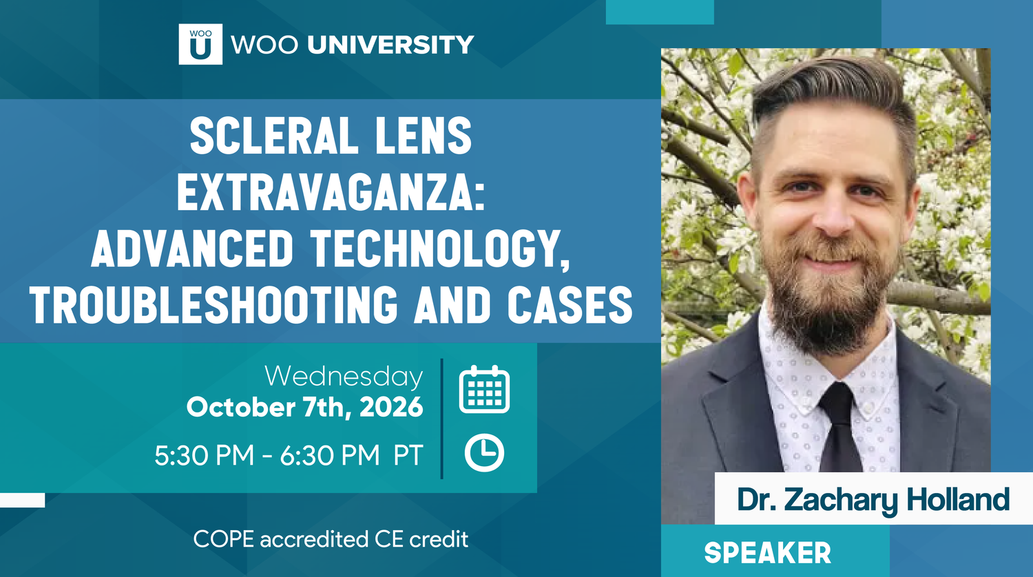
Scleral Lens Extravaganza! Advanced Technology, Troubleshooting and Cases
Featuring Dr. Zac Holland
Successful scleral lens fitting requires more than achieving adequate central clearance. In our recent Woo U CE event, Dr. Zac Holland presented Scleral Lens Extravaganza: Advanced Technology, Troubleshooting and Cases, exploring complex fitting challenges, advanced diagnostic technologies, and customized lens designs to improve patient comfort and visual outcomes.
From troubleshooting landing zone alignment to incorporating profilometry and higher-order aberration (HOA) correction, Dr. Holland shared practical clinical insights for managing challenging scleral lens fits.
Key Clinical Takeaways
1. Evaluate the entire fit, not just central clearance.
A successful scleral lens fit requires careful assessment of central clearance, limbal clearance, landing zone alignment, lens rotation, and centration. Each contributes to lens stability, comfort, and ocular health over time.
2. Landing zone alignment is critical.
An overly tight landing zone may cause blanching, compression rings, and discomfort, particularly toward the end of the day. Conversely, an excessively loose landing zone may contribute to lens movement, decentration, and fogging. Identifying the location and pattern of misalignment helps determine whether toric, quadrant-specific, or freeform haptic adjustments are appropriate.
3. Identify the underlying cause of midday fogging.
Midday fogging can have multiple contributing factors, including debris accumulating within the tear reservoir and issues with surface wettability. Lens alignment, filling solutions, ocular surface conditions, and lens surface characteristics should all be considered before making adjustments.
4. Don't overlook limbal clearance.
Insufficient limbal clearance may contribute to epithelial changes, including microcystic changes and staining, and may be associated with patient discomfort or end-of-day dryness. Evaluating limbal clearance after the lens has settled is important, particularly when diameter or decentration may be affecting the fit.
5. Use profilometry to guide complex fits.
Profilometry provides detailed corneoscleral elevation data that can help clinicians evaluate scleral asymmetry, anticipate lens rotation, and customize the landing zone. For challenging fits, this technology may reduce reliance on traditional trial-and-error adjustments.
6. Customize more than the haptic when necessary.
Localized elevation differences may require adjustments to more than the landing zone. Dr. Holland demonstrated how independently modifying midperipheral, limbal, and landing zone curves can help address specific anatomical variations and fitting complications.
7. Excellent visual acuity doesn't always mean excellent visual quality.
Patients may achieve 20/20 vision yet continue to experience halos, glare, or other visual disturbances. Wavefront-guided scleral lenses may help address residual higher-order aberrations that conventional optical correction does not fully resolve.
In one keratoconus case presented, HOA RMS decreased from 1.12 to 0.63 µm OD and from 1.25 to 0.63 µm OS, alongside significant improvement in the patient's visual complaints.
8. Establish a stable fit before incorporating advanced optics.
Lens centration and rotational stability are particularly important when applying HOA correction. Dr. Holland's cases highlighted the importance of addressing complex fitting challenges, including those guided by profilometry, before introducing advanced optical customization.
The Takeaway
When a scleral lens isn't performing as expected, identifying the underlying cause is essential before making adjustments. A systematic approach to evaluating the fit, combined with advanced diagnostic tools and increasingly customizable lens designs, can help clinicians better address patient comfort, lens stability, and visual quality.

Woo’s Wisdom: From Lesions to Lids — Practical Applications of Chemical Cautery in Optometry
Featuring Dr. Nathan Lighthizer
Recognizing eyelid lesions, distinguishing benign findings from potentially concerning features, and selecting appropriate management strategies are important aspects of clinical practice. In our recent Woo U CE event, Dr. Nathan Lighthizer presented From Lesions to Lids: Practical Applications of Chemical Cautery in Optometry, covering lesion assessment, common eyelid conditions, and treatment considerations.
Key Clinical Takeaways
1. Recognize suspicious features using the ABCDs.
Dr. Lighthizer discussed key characteristics to evaluate when assessing a lesion for potentially concerning features, including:
A – Asymmetry: Is the lesion asymmetrical?
B – Blood vessels: Are there abnormal or prominent vascular features?
C – Borders: Are the borders irregular?
D – Different colors: Is the lesion multicolored or unevenly pigmented?
He also emphasized monitoring for changes in size, color, and overall diameter over time.
2. Take a thorough lesion history.
A proper lesion workup involves more than evaluating its appearance. Important questions include:
How long has the lesion been present?
Has it changed in size, color, or appearance?
Is it bothersome, irritated, or bleeding?
Does the patient have a personal history of cancer?
These details help guide clinical assessment and determine whether further evaluation or referral is warranted.
3. Understand the presentation of common benign eyelid lesions.
The presentation explored the clinical features, differential diagnoses, and management considerations of several common eyelid lesions, including:
Verruca vulgaris
Squamous cell papilloma
Seborrheic keratosis
Cyst of Zeis
Cyst of Moll
Epidermal inclusion cyst
Pyogenic granuloma
Cherry hemangioma
Molluscum contagiosum
Eyelid nevus
Understanding how these lesions present can help clinicians recognize common findings, distinguish between potential diagnoses, and determine appropriate next steps.
4. Explore treatment options, including chemical cautery.
Dr. Lighthizer discussed treatment approaches for various eyelid lesions, including the clinical application of chemical cautery. Through case studies, he demonstrated its use in selected cases and shared practical considerations for lesion management.
The Takeaway
A thorough eyelid lesion evaluation involves recognizing clinical features, assessing changes over time, obtaining a relevant patient history, and understanding when treatment or further evaluation is appropriate. Familiarity with common benign lesions and treatment options, including chemical cautery, can help support informed clinical decision-making.
Thank you to Dr. Nathan Lighthizer for sharing his clinical knowledge and practical approach to eyelid lesion evaluation and management.
Watch the full presentation and access the handouts at WooU.org under Past Events.
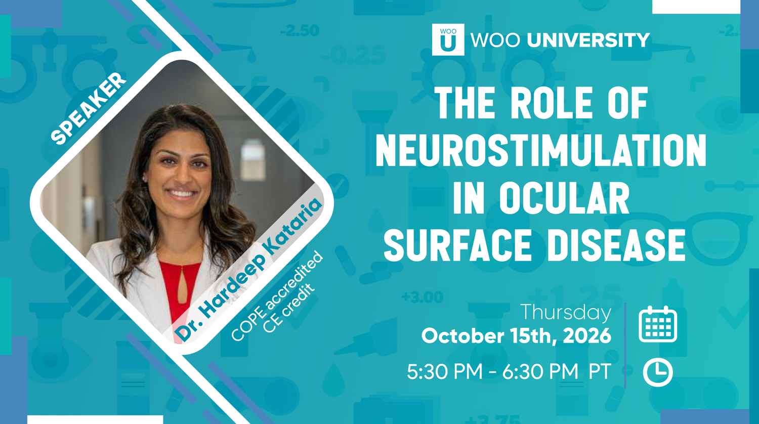
Woo’s Wisdom: The Role of Neurostimulation in Ocular Surface Disease
Featuring Dr. Hardeep Kataria
Dry eye disease involves more than tear film quantity and quality. Sensory signaling and neural regulation also play important roles in maintaining ocular surface homeostasis. In our recent Woo U CE event, Dr. Hardeep Kataria presented The Role of Neurostimulation in Ocular Surface Disease, exploring the relationship between neural pathways, tear production, and emerging treatment approaches.
Key Clinical Takeaways
1. Understand neuromodulation and neurostimulation.
Dr. Kataria began by defining neuromodulation and neurostimulation, explaining how sensory stimulation interacts with neural pathways and reflex mechanisms involved in ocular surface function.
2. Recognize the role of neural regulation in dry eye disease.
Dry eye disease is not simply a problem of insufficient tear volume. Sensory input and neural regulation influence tear secretion and ocular surface homeostasis. Understanding these mechanisms provides a broader perspective on the disease and its treatment.
3. Explore different neurostimulation modalities.
The presentation reviewed several approaches to neurostimulation for ocular surface disease, including:
Intranasal stimulation: Mechanical and chemical approaches
Extranasal stimulation
Corneal stimulation
Each modality involves different approaches to activating sensory pathways involved in tear film regulation.
4. Consider patient selection and practical application.
Dr. Kataria shared clinical pearls, tips for administration, and considerations for identifying which dry eye patients may benefit from different neurostimulation approaches. She also discussed how these therapies may be incorporated into a broader dry eye management plan.
5. Look toward the future of neural homeostasis.
The discussion concluded with the evolving role of neural regulation in ocular surface disease, with a focus on restoring a balanced neural-lacrimal feedback loop. Understanding this relationship may help shape future approaches to dry eye treatment.
The Takeaway
Neurostimulation expands the way clinicians think about dry eye disease by addressing the sensory and neural pathways involved in tear regulation, rather than focusing solely on the tear film itself. Understanding the available modalities, patient considerations, and their place within treatment strategies provides another perspective on managing ocular surface disease.
Thank you to Dr. Hardeep Kataria for sharing her expertise and insights into the evolving role of neurostimulation in dry eye management.
Watch the full presentation and access the handouts at WooU.org under Past Events.
