Woo U Wisdom: A Comprehensive Guide to Glaucoma Care - Treatment Approaches and Managing Comorbidities

Open or Closed: How We Treat Glaucoma with Dr. Mitch Ibach
This webinar provided a practical, clinically focused review of modern glaucoma management, from first-line treatment options for open-angle glaucoma to urgent management of acute angle closure. Dr. Ibach compared topical drops, SLT, drug delivery, and MIGS through the lens of efficacy, patient adherence, cost, safety, and long-term disease control. Dr. Ibach also emphasized the importance of gonioscopy, individualized treatment planning, and recognizing secondary causes of elevated IOP.
Key Takeaways:
Prostaglandin analogs remain foundational: Latanoprost continues to have strong evidence supporting its role in reducing visual field progression in glaucoma patients. Newer options, including preservative-free latanoprost, may improve tolerability for patients with ocular surface disease while maintaining similar efficacy.
SLT is a strong first-line treatment option: In open-angle glaucoma patients, SLT often lowers IOP similarly to a prostaglandin analog while avoiding many compliance issues associated with drops.
Drug delivery may significantly improve adherence: Sustained-release options such as Durysta and iDose offer the potential to reduce dependence on daily topical medications, minimize preservative exposure, and improve long-term treatment consistency.
MIGS plays an important role alongside cataract surgery: For patients with mild to moderate glaucoma undergoing cataract surgery, MIGS can provide additional IOP lowering with a favorable safety profile and minimal added recovery time.
Gonioscopy remains critical in glaucoma care: Proper angle assessment is essential for accurately differentiating open-angle glaucoma, narrow angles, plateau iris, and secondary causes of elevated IOP.
Angle closure requires rapid recognition and intervention: Acute angle closure is an ocular emergency. Management focuses on quickly lowering IOP, identifying the underlying mechanism, and developing an appropriate long-term surgical or medical plan.
Clear lens extraction may be favored in select angle-closure patients: Based on the EAGLE study, clear lens extraction may provide better long-term outcomes than LPI alone in certain patients over age 50 with primary angle closure or primary angle closure glaucoma.
Elevated IOP can have many different mechanisms: Uveitic glaucoma, neovascular glaucoma, steroid response, retained viscoelastic, hyphema, medication-induced angle closure, and aqueous misdirection all require different clinical reasoning and management approaches.
Optimizing Glaucoma Care: Managing the Comorbidities with Dr. Jessica Steen
This lecture focused on the complexities of modern glaucoma care beyond simply lowering intraocular pressure. Dr. Steen emphasized individualized risk assessment, strategic use of OCT and visual field testing, interpretation of glaucoma imaging in the presence of retinal comorbidities, and the growing impact of ocular surface disease on long-term treatment adherence. The session also highlighted how retinal pathology, high myopia, anti-VEGF therapy, and systemic disease can significantly complicate glaucoma diagnosis and progression analysis.
Key Takeaways:
Intraocular pressure is only one piece of the puzzle: Elevated IOP remains the most significant modifiable risk factor for glaucoma progression, but “normal” pressure is highly individualized. Patients can develop glaucoma at statistically normal pressures, while others tolerate higher pressures without damage.
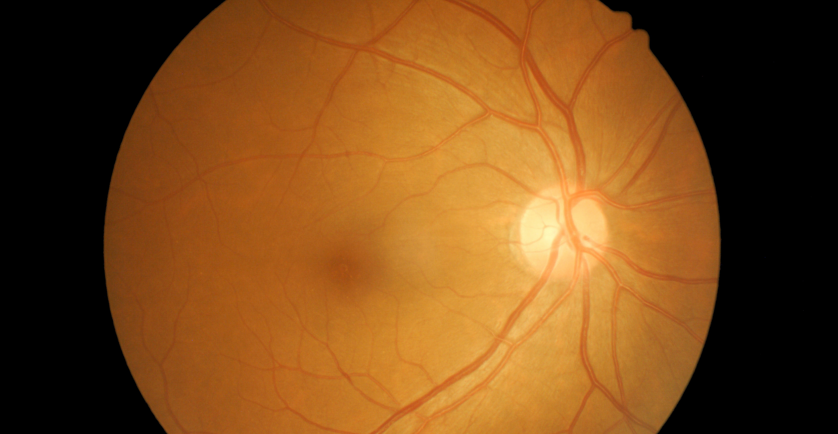
Glaucoma diagnosis starts with the optic nerve, not the tonometer: Risk assessment should include optic nerve evaluation, pachymetry, gonioscopy, imaging, visual fields, systemic history, refractive status, and vascular risk factors rather than relying on IOP alone.
Testing frequency should be individualized: Rather than using a “one-size-fits-all” schedule, glaucoma monitoring should be tailored to disease severity, progression risk, and patient-specific findings. Clustering OCTs and visual fields earlier in the disease process may improve detection of progression.
Gonioscopy remains essential in modern glaucoma care: Even in pseudophakic patients, periodic gonioscopy is important to assess angle anatomy, pigment, peripheral anterior synechiae, MIGS positioning, and secondary glaucoma risk factors.
Ocular surface disease is extremely common in glaucoma patients: Glaucoma drops do not necessarily “cause” dry eye, but they frequently worsen underlying ocular surface disease. Preservatives, chronic medication exposure, autoimmune disease, and inflammation all contribute to reduced comfort and poorer adherence.
Patient comfort directly affects medication adherence: Addressing ocular surface disease early can improve long-term compliance with glaucoma therapy. The presentation emphasized using validated symptom questionnaires and proactively treating even minimally symptomatic patients.
Preservative-free glaucoma therapies continue to expand: Newer preservative-free prostaglandin analogs and delivery systems may improve tolerability while maintaining strong IOP-lowering efficacy, especially in patients with chronic ocular surface disease.
Glaucoma patients often have overlapping retinal disease: Conditions such as retinal vein occlusions, high myopia, AMD, hydroxychloroquine toxicity, epiretinal membranes, and myopic degeneration can complicate interpretation of OCTs and visual fields. Clinicians must always ask whether structural or functional change is truly glaucomatous.
OCT interpretation requires careful clinical context: The session emphasized looking beyond color-coded printouts and instead evaluating where the pathology is located anatomically. Understanding retinal, choroidal, and optic nerve pathology is critical when interpreting imaging in glaucoma patients.
High myopia significantly increases glaucoma complexity: Highly myopic eyes are more difficult to monitor structurally and functionally, and these patients also carry increased risk for progressive myopic macular degeneration and choroidal neovascularization.
Anti-VEGF injections can impact glaucoma risk: Intravitreal injections cause significant transient IOP spikes and may contribute to long-term trabecular dysfunction, particularly in patients receiving frequent injections over many years. Glaucoma patients receiving retinal injections require careful long-term monitoring.
Managing glaucoma requires a whole-patient approach: The presentation repeatedly emphasized that glaucoma care extends beyond simply lowering pressure. Successful management requires understanding systemic disease, retinal comorbidities, medication burden, patient quality of life, and individualized risk over time.
Glaucoma management continues to evolve, requiring clinicians to balance evidence-based treatment strategies with the unique needs of each patient. As highlighted by Dr. Mitch Ibach, successful treatment begins with accurate diagnosis, careful angle assessment, and selecting therapies that optimize intraocular pressure control while supporting long-term adherence. Advances in laser therapy, sustained drug delivery, MIGS, and surgical interventions have expanded the options available for both open-angle and angle-closure disease.
At the same time, Dr. Jessica Steen emphasized that effective glaucoma care extends well beyond pressure reduction alone. Retinal comorbidities, ocular surface disease, high myopia, systemic health factors, and treatment burden can all influence disease progression, diagnostic interpretation, and patient outcomes. A comprehensive approach that integrates structural and functional testing, individualized monitoring schedules, and proactive management of comorbid conditions is essential for preserving vision and maintaining quality of life.
Together, these presentations reinforce an important message: optimizing glaucoma care requires both precision in treatment selection and a holistic understanding of the patient. By combining modern therapeutic advances with thoughtful, patient-centered management, clinicians can better navigate the complexities of glaucoma and improve long-term outcomes for those living with this chronic disease.